Altris for Geographic Atrophy Research
Assessment of imaging features associated with GA (RUO)
Book Intro
Altris IMS is an FDA-cleared (510(k)) ophthalmic data-management system for securely importing, storing, and managing OCT data. Intended for use by eye-care professionals in clinical workflows to manage patient imaging data and generate clinical reports & support research workflows by enabling:
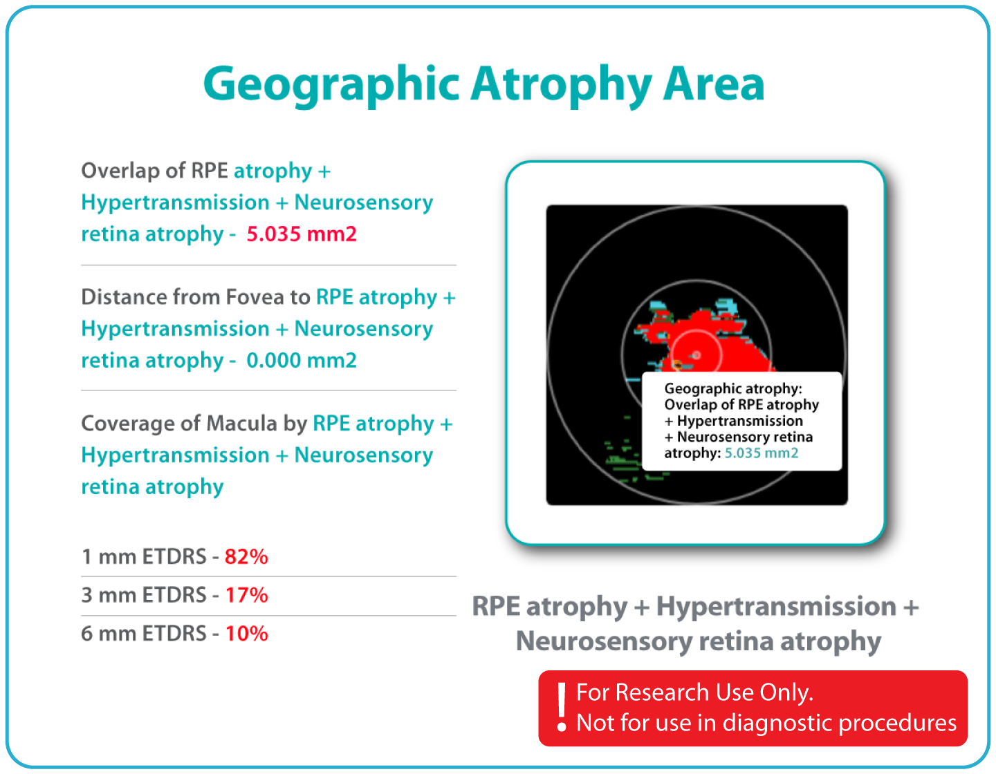
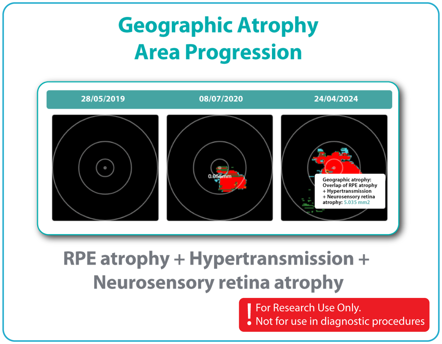
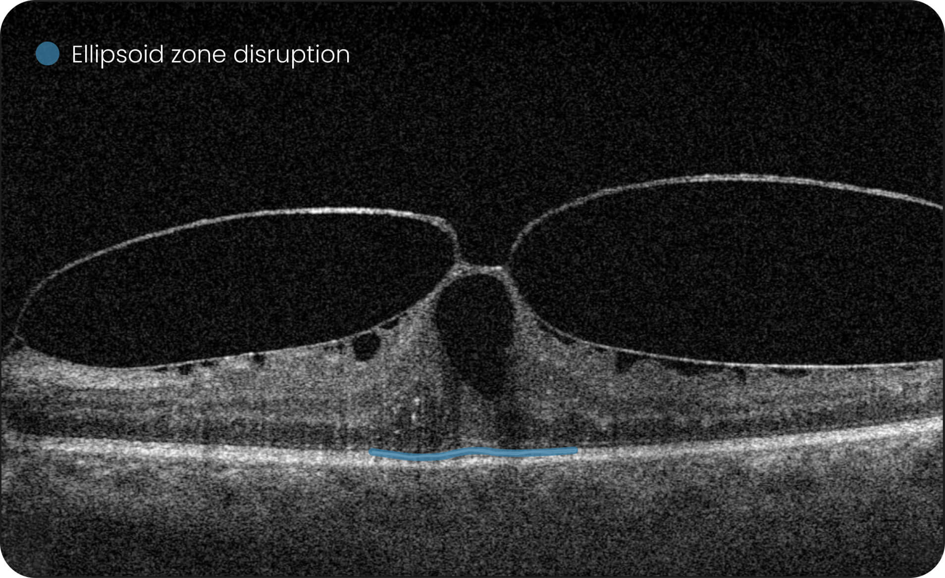
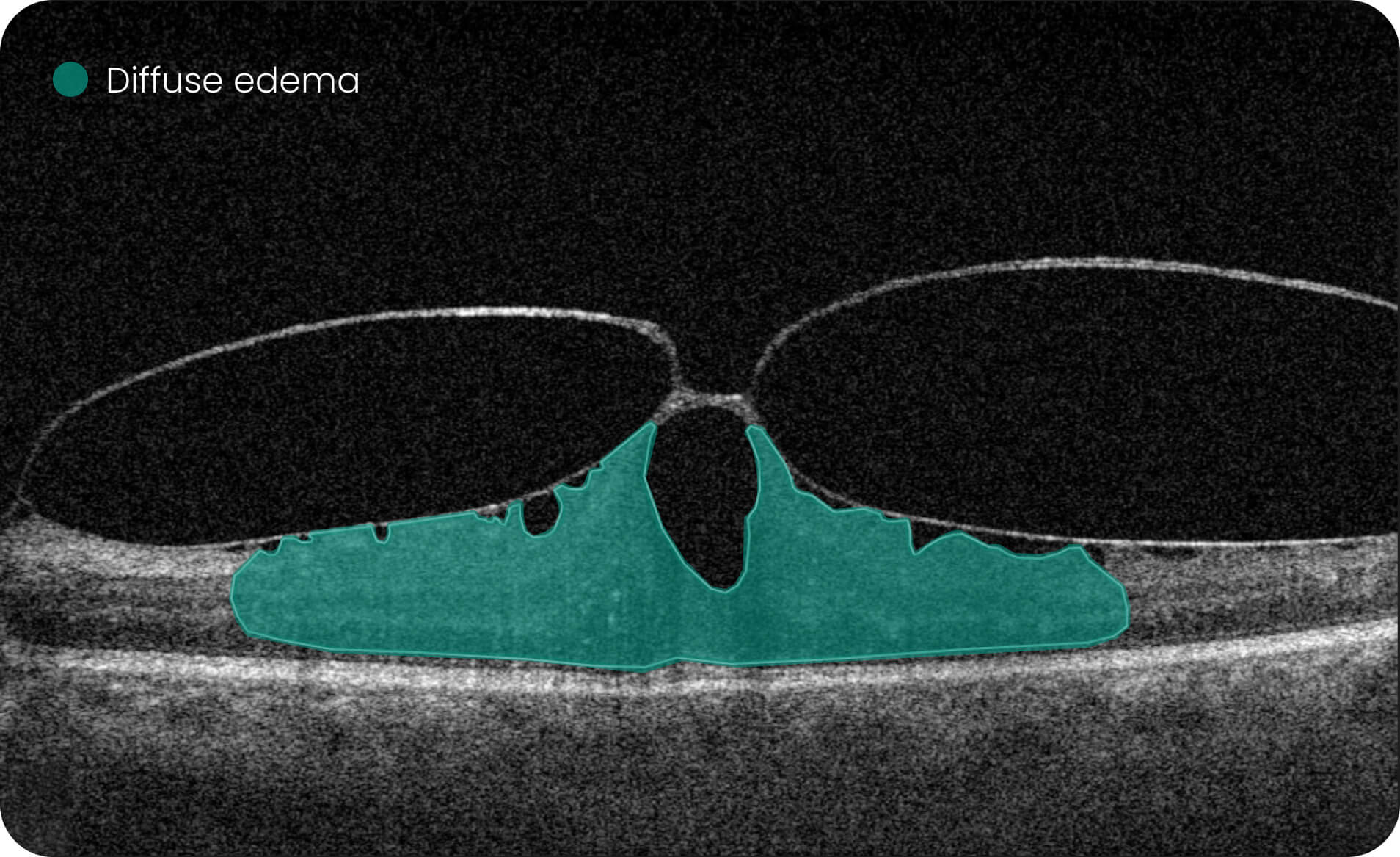
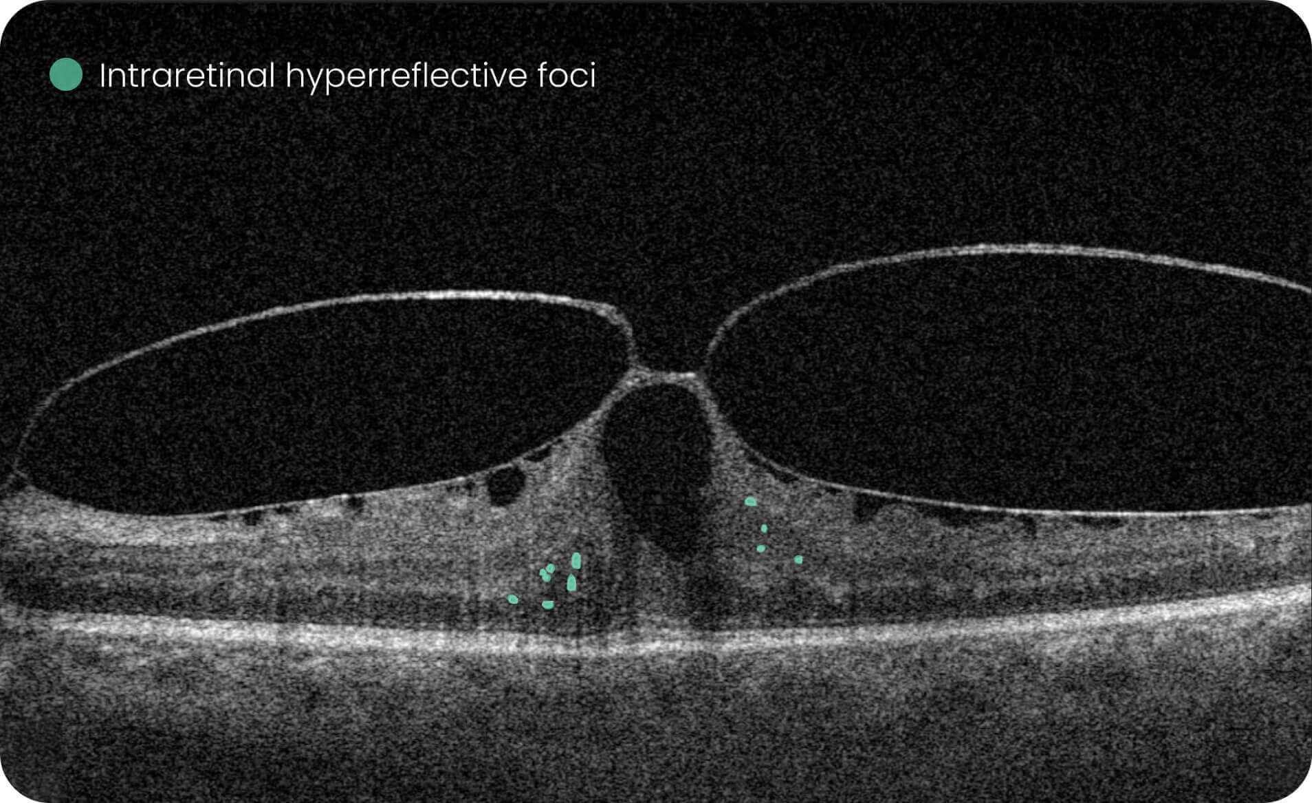
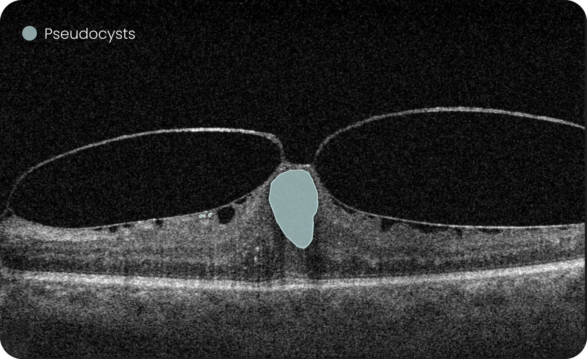
Characterization and visualization of OCT features commonly studied in Geographic Atrophy (GA), including hypertransmission, RPE atrophy, neurosensory retinal atrophy, and EZ changes.
Estimation of GA-related feature areas using combinations of imaging characteristics.
Longitudinal review of feature changes across multiple examinations, expressed in linear or percentage-based measurements.
For Research Use Only. Not for diagnostic or clinical decision-making.


Platform overview
Only practical features for eye care specialists and clinical research
The security of patients’ data is our top priority: we are GDPR compliant, all data is encrypted, CE-certified, and FDA-cleared (510k).
 Alisdair Buchanan
Optometry Owner, UK
Alisdair Buchanan
Optometry Owner, UK

 Clara Pereira
Optometrist, Portugal
Clara Pereira
Optometrist, Portugal
 Jeff Sciberras
Optometry Owner, Canada
Jeff Sciberras
Optometry Owner, Canada
The GA Progression feature provides tools for research-oriented visualization and comparison of OCT imaging data across multiple visits. Users can review changes in areas associated with geographic atrophy (GA) and related imaging features through percentage-based displays, maps, and graphs.
-
Efficient estimations of GA-associated areas designed to support research-focused image review workflows.
-
Quantitative, image-derived measurements of GA-related features and associated biomarkers for research analyses.
-
Visualization of spatial relationships, including the estimated distance between GA-associated regions and the foveal area, as well as macular involvement.
-
Support for research dataset stratification through calculations based on the overlap of multiple imaging-feature sets. For Research Use Only. Not for diagnostic or clinical decision-making.
How Altris Works?
And what's the value of using Altris for Geographic Atrophy Research?

Altris is a web platform developed by professionals with expertise in retinal imaging. We’ve collected a large number of OCT scans, and our team has manually annotated thousands of them to develop the Altris system, an artificial intelligence geographic atrophy research USA platform, which can:

Formats
DICOM format will help you to extract maximum information. However, the system works with all
data formats, such as jpg, and png

OCT equipment
Altris AI is vendor-neutral. We work with all the OCT equipment producers

OCT reports
We create comprehensible OCT reports for eye care researchers
For Pharma
For innovative approach in Pharma
Contact usWhat you get with Altris:
- Centralized OCT Data Management
- Historical Data Analysis
- Vendor-neutral analysis of OCT scans (8 manufacturers)
- Data Security & Compliance
- Additional capabilities for research
- 40+retinal biomarkers studied in research across 30+ retinal conditions. For Research Use Only. Not for diagnostic procedures.
- Quantitative exploration of 40+ biomarkers for Research Use Only. Not for diagnostic procedures.
- Reports
For Eye Care
IMS for Ophthalmology and Optometry
Contact usWhat you get with Altris:
- Centralized OCT Data Management
- Vendor-Neutral OCT Compatibility (8 manufacturers)
- Historical OCT Data Analysis
- Seamless Clinical Workflow Integration
- Informative OCT reports
- 40+retinal biomarkers studied in research across 30+ retinal conditions. For Research Use Only. Not for diagnostic procedures.
- Quantitative exploration of 40+biomarkers for Research Use Only. Not for diagnostic procedures.